Gretchen Anderson came to The University of Alabama to work with animals and pursue veterinary studies, but she ended up with human bones on her hands.
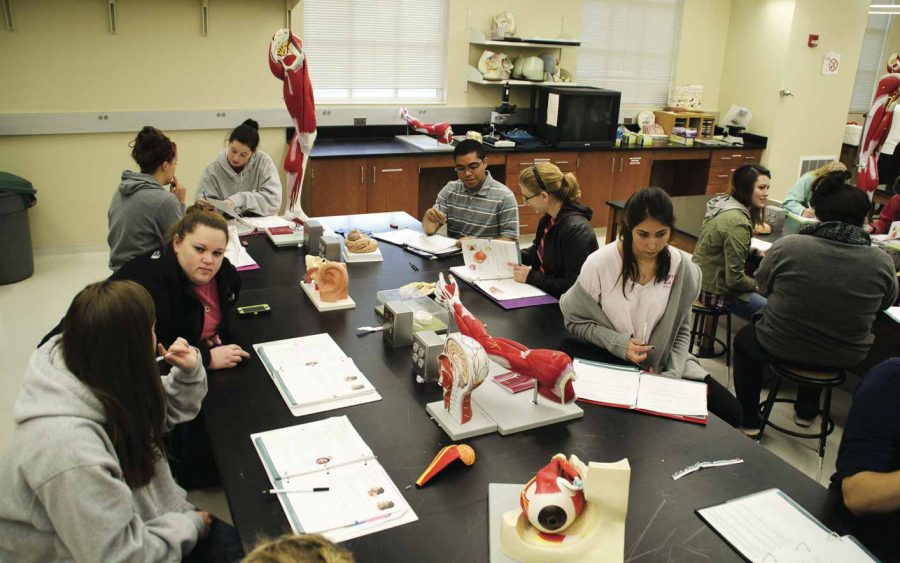
As the lead graduate teaching assistant for Anatomy and Physiology 215, Anderson uses human specimens and synthetic models to teach her students.
“In the lab we’re just trying to give them a little bit more hands on, and it’s a lot more anatomy than physiology in the lab,” Anderson said. “We show them models or individual specimens that they can look at and touch to go through the skeletal system, muscular system, special senses, nervous system, stuff like that.”
Anderson said she believes that while synthetic bone models are increasingly accurate, it is important for students to see real life examples of diversity between specimens. She said the use of synthetic models is becoming more prominent than human specimens now.
“We use mostly synthetic. The only animal parts we use are sheep brains and cow eyes. We used to dissect cats in the class to do muscles, but this year we’ve switched over to using synthetic muscle models, which is good and bad,” Anderson said. “It’s bad because they’re missing out on the dissection experience. It’s good because instead of learning cat muscles, they’re learning human muscles, which is much more relevant for them.”
Brook Fluker, an assistant professor in biological sciences, said human bones provide students with details that models may not include. This opportunity is particularly beneficial to students interested in health care.
“For our bone labs most of what we’re doing is, number one, what do these bones look like without skin and muscle on top. We have a femur sectioned in half where students can see where the bone marrow is created,” Fluker said. “They get a void view to see those types of structures in the bones. This may be the only anatomy class they have, so we have to try to teach as much as possible.”
Anderson also said real examples better translate anomalies in bone structure. The human bones at the University include long bones, pelvises and a few skulls.
“You can look at a bone model all you want, but when you actually see a real bone you realize that it’s called spongy bone because it’s actually porous,” Anderson said.
With improving technology, lab materials are becoming increasingly synthetic. Students now interact with mostly plastic structures while learning the body. These plastic models include a full skeleton that has even been named by the department.
“We do have a full fake skeleton,” Fluker said. “His name is Bob. We also have a disarticulated set, a Bob-in-a-box.”
Anderson said times are shifting away from dissection and human specimens. She has not had any student controversy about using the specimens, though, even as they become less popular.
“The bones that we use have all been in the lab for a very long time. They’re old and they’re from a previous time where they didn’t really have these synthetic models as much,” Anderson said. “When you tell them that’s a real bone, people are like ‘Ew, really? I want to touch it.’ I’ve never really had anyone that has said that it’s not right.”
Fluker said any human bones would not have been taken without consent. People can choose to donate their bodies to scientific companies that allow schools to purchase them for research and teaching.
“Really what they’re looking at, though, is just a shell of what it was. It’s just the minerals. A living bone is very much alive, but these are just the shells. And, to the best of my knowledge, most of the real bones provided by scientific companies are the product of someone donating their body to science,” Fluker said.